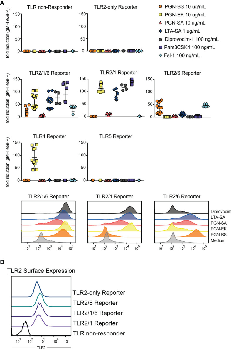
Figure 5.

TLR2 stimulatory capacity of different cell wall components. (A) Activation of indicated reporter cells by the microbial components peptidoglycan (PGN), isolated from the gram-positive B. subtilis (PGN-BS) or S. aureus (PGN-SA) or the gram-negative E. coli K12 (PGN-EK), purified lipoteichoic acid from S. aureus (LTA-SA) and the synthetic ligands Diprovocim-1 (TLR2/1), Pam3CSK4 (TLR2/1) and Fsl-1 (TLR2/6) (n = 8, each experiment performed in duplicates). Exemplary histograms of eGFP expression are shown for TLR2/1/6, TLR2/1 and TLR2/6 reporter cells. (B) TLR2 surface expression on different TLR reporter cell lines was compared by staining with an AF647-coupled anti-TLR2 antibody.
