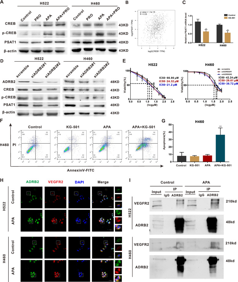
Fig. 5. The mechanism of apatinib resistance in NSCLC cells.
A H522 and H460 cells were treated with or without apatinib (20 μM) and propranolol (50 μM) for 48 h. The relative protein level of CREB, p-CREB, PSAT1 was detected through Western blot. B A positive correlation between CREB and PSAT1 expression in lung cancer according to TCGA database. C qRT-PCR analysis of PSAT1 mRNA levels in H522 and H460 cells after KG-501 treatment. D Western blot analysis of CREB, p-CREB, PSAT1 expression in H522 and H460 cells after ADRB2 knockdown. E H522 and H460 cells were transfected with indicated siRNAs for 48 h, and the samples were exposed to different concentrations of apatinib. IC50 of apatinib was measured with CCK8 assay. F H460 cells were treated with/without apatinib (20 μM) and KG-501 (25 μM) for 48 h. The apoptosis index was detected by flow cytometry. G The percentage of apoptotic cells in H522 and H460 cells. H Representative co-localization images stained with ADRB2 (green) and VEGFR2 (red) in H522 and H460 cells. I Anti-ADRB2 antibodies immunoprecipitated endogenous ADRB2 and Co-IP VEGFR2 by Western blot analysis. (**P < 0.01, compared with vehicle).

