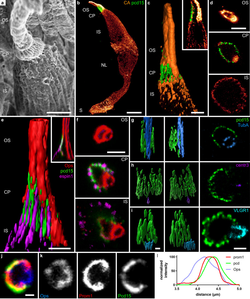
Fig. 2. Calyceal C-shaped palisade around the cone cilium.
a Scanning electron microscopy of a primate cone showing the outer and inner segment as well as the calyceal process around the base of the outer segment. 3D reconstructions (b, c, e g–i), reconstructed coronal slices (c and e inserts) and optical sections (d, f, last image of g–i, and j–k) of cone photoreceptors immunolabeled for the R/G cone opsin (red in a, d–g; blue in l), espin 1 (magenta e, f), Protocadherin15 (green), cone arrestin (orange in b–d), prominin 1 (red in j), acetylated alpha tubulin (blue in g), centrin 3 (purple in h), and VLGR1 (cyan in i). l normalized intensity across the cellular border. The Pcd15 staining is located in a position external to prominin 1. Numerical data in supplementary data 2. OS outer segment, CP calyceal processes, IS inner segment, NL nuclear layer, S synapse, arrowhead: pcd15 staining expression around the periciliary membrane at the base of the cilia. Scale bars represent 2 µm in (a, c, d) and 1 µm in (e–j), and 10 µm in (b).

