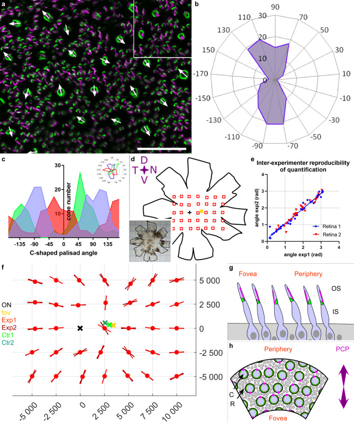
Fig. 5. Planar polarity of cone photoreceptors in the macaque retina.
a Flat-mounted retina immunostained for protocadherin 15 (pcd15, green) and acetylated tubulin alpha (alpha-Tub, magenta), showing the directions vectors for all the cones in the image. Each cone vector is defined by the C-shaped symmetric axis of the Pcd15 staining and the direction toward the opening of the C-shape defined by the acetylated tubulin alpha staining, as illustrated in the inset. (b) Polar plot of the vectors on a retinal image (0.25 mm²) showing a clear alignment along a preferential planar axis. c Graphs representing the distributions of vector directions for 3 independent retinas, showing that all retinal samples had two peaks in opposite directions (numerical data in supplementary data 5). d–f Cone vector directions for the primate retina are shown in d. The planar axes are illustrated on the matrix (f) for all sample points located on the schematic representation of the retina (d). For each location, two experimenters independently placed individual cone vectors and calculated the mean planar axis. e Mean planar axis angle, demonstrating the reproducibility of the technique. (numerical data in supplementary data 6) f Graph providing the angles of the planar axis obtained for macaque 1 and for each observed retinal location in red (different hues for the datasets of different experimenters). Each planar axis was calculated from a minimum of 50 individual cone vector angles. The optic nerve, fovea, and both reconstructed centers (from the two different datasets) are represented by black, yellow, and green crosses, respectively. (macaque n2 in supplementary figure, and numerical data in supplementary data 7). g, h Schematic representation of the planar polarity organization discovered (g: view as a retinal radial section. Cones appear tilted toward the fovea. h: view as a flat-mounted retina. C cones, R rods, Green calyceal processes, magenta cilia). See Supplementary figure for comparison of macaque 1 and 2.

