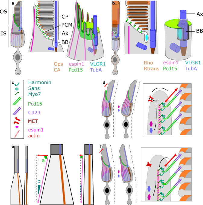
Fig. 7. Schematic diagram of the distribution of USH proteins, other mechanical proteins, and cilium-associated proteins in rod and cone photoreceptors.
(a: cone, b rod). Schematic reconstruction of cone 3D inner/outer segment junction structure, showing: full photoreceptor view, a detailed transverse view, and a 3D reconstitution of the apical structure of the inner segment (outer segment remove for visual clarity). e Difference between cones and rods in inner-to-outer segment diameter ratio, leading to different effects of identical shortenings of the inner segment. d, f hypothesis for outer segment tilting leading to a change in inner segment alignment, c legend of the symbols are used in panels d and f. Ax axoneme, BB basal body, CP calyceal process, PCM periciliary membrane, OS outer segment, IS inner segment.

