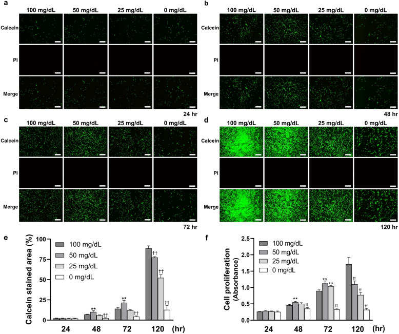
Figure 1.
Low glucose concentrations affect the cell proliferation on human gingival fibroblast (HGnFs). (a, b, c and d) HGnFs stained by calcein-AM and PI were photographed under a fluorescence microscope after incubation for 24, 48, 72 and 120 h. (e) The data of viable and dead staining are presented as the percentage of calcein stained area. (f) Cell proliferation was measured after incubation for 24, 48, 72 and 120 h. (Scale bars: 500 μm; A significant increase compared with the control was described as **P < 0.01; A significant decrease compared with the control was described as ††P < 0.01).

