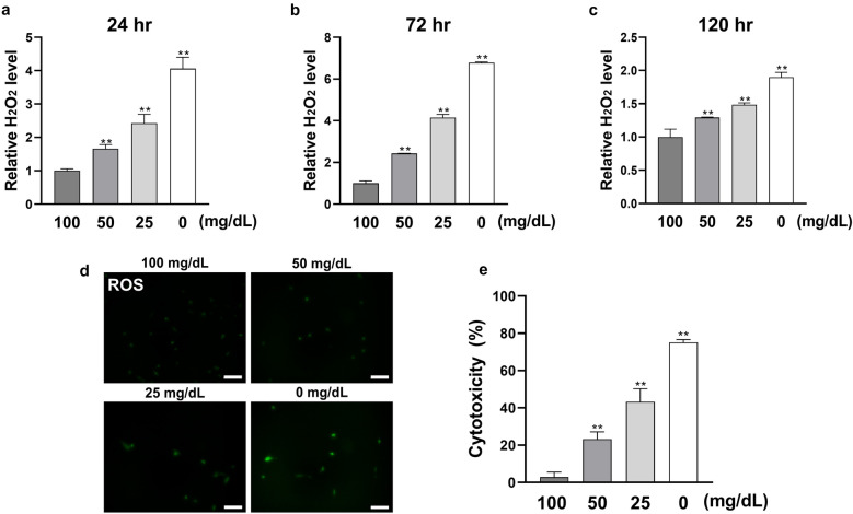
Figure 3.
Low glucose concentrations enhance cellular stress level of HGnFs. (a, b and c) The level of ROS was examined using hydrogen peroxide (H2O2) detection kit. After 24, 72 and 120 h of incubation, the supernatant was collected and the level of H2O2 was measured by a microplate reader, and the results was modified relative to the control group. (d) The ROS stained cells were photographed by a fluorescence microscope after incubation for 120 h. (e) Cytotoxicity (LDH level) was determined after 24 h of incubation. (Scale bars: 100 μm; A significant increase compared with the control was described as **P < 0.01).

