Figure 6.
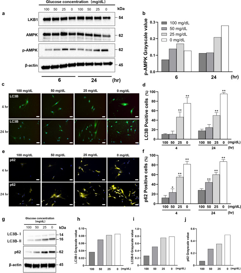
Low glucose concentrations activate the LKB1-AMPK signaling pathway and induce autophagy. (a and b) The protein expression of LKB1-AMPK signaling pathway was evaluated (a) using western blotting analysis and quantified using ImageJ (b). Western blot analysis was performed on protein extracts of these cells with antibodies against the indicated proteins with β-actin as a loading control. The samples derive from the same experiment and those gels/blots were processed in parallel. Uncropped blots for this experiment are presented in supplementary file. The expression levels of LKB1, AMPK and p-AMPK were measured by densitometric analysis using ImageJ. (c–j) The synthesis of autophagy-related proteins (LC3B and p62) by HGnFs under low glucose concentrations. HGnFs were photographed after being incubated with fluorescently labeled secondary antibody and stained with DAPI (c and e). The data of the synthesis of LC3B and p62 are presented (d and f) as the ratio of the positive cells. The expression of LC3B and p62 were examined using western blotting (g) and analyzed using ImageJ (h, i, and j). Western blot analysis was performed on protein extracts of these cells with antibodies against the indicated proteins with β-actin as a loading control. The samples derive from the same experiment and those gels/blots were processed in parallel. Uncropped blots for this experiment are presented in supplementary file. The expression levels of p62, LC3B-I and LC3B-II were measured by densitometric analysis using ImageJ. (Scale bars: 100 μm; A significant increase compared with the control was described as *P < 0.05, **P < 0.01).

