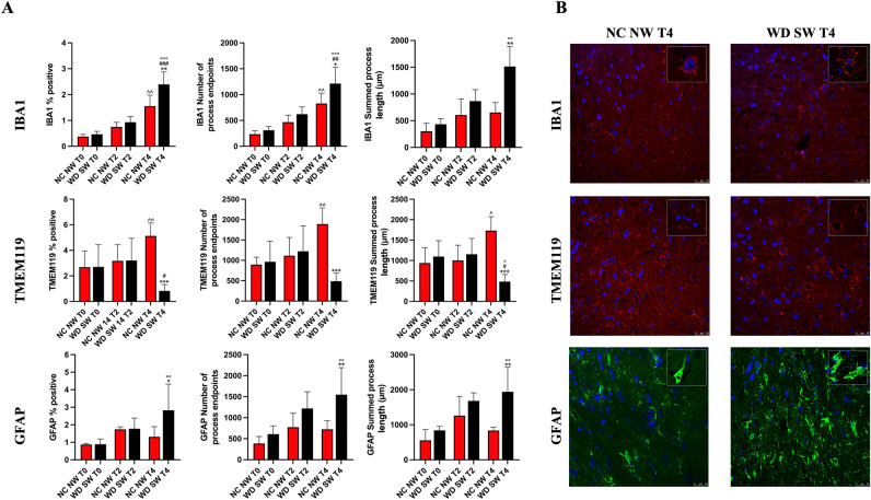
Figure 4.
Immunofluorescence staining for IBA1 and TMEM119 in microglia and for GFAP in astrocytes. (A) The results are expressed as mean ± S.D. NC NW: Normal Chow Normal Water; WD SW: Western Diet Sugar Water. T0: 0 weeks; T2: 8 weeks; T4: 28 weeks. IBA1: Ionized calcium-binding adaptor molecule 1, TMEM119: Transmembrane protein 119, GFAP: Glial fibrillary acidic protein. *: p < 0.05, **: p < 0.01, ***: p < 0.001 vs NC NW T4; #: p < 0.05, ##: p < 0.01, ###: p < 0.001 vs WD SW T2, °: p < 0.05, °°: p < 0.01, °°°: p < 0.001 vs WD SW T0; ^: p < 0.05, ^^: p < 0.01 vs NC NW T0. (B) Representative confocal images of IBA1 positive microglia (above), TMEM119 positive microglia (middle), GFAP positive astrocytes (below) and magnified illustrations of microglia and astrocytes morphology.

