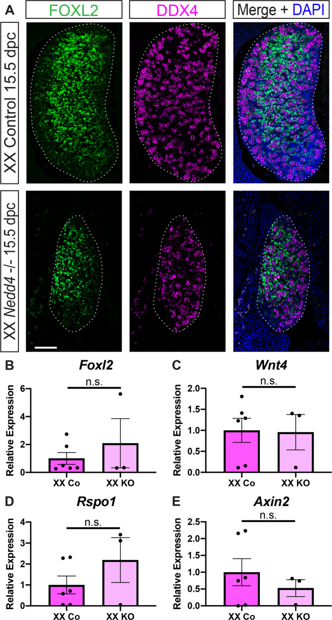
Fig. 3. Normal ovarian somatic cell development in XX Nedd4-/- mice.
A Section immunofluorescence on 15.5 dpc XX Nedd4-/- embryos alongside XX littermate controls stained for ovarian somatic cells markers FOXL2 (green) and germ cell marker DDX4 (magenta). The anterior pole of each gonad is positioned at the top of each panel. Gonads are denoted by a white dotted line. Scale bars = 100 μm. (B–E) RT-qPCR analyses of Foxl2 (B), Wnt4 (C), Rspo1 (D) and Axin2 (E) expression at 14.5 dpc on XX Nedd4-/- gonads (KO) (n = 3) and XX controls (n = 6). Values are expressed relative to the XX controls. Mean ± SEM; t-test, n.s. not significant.

