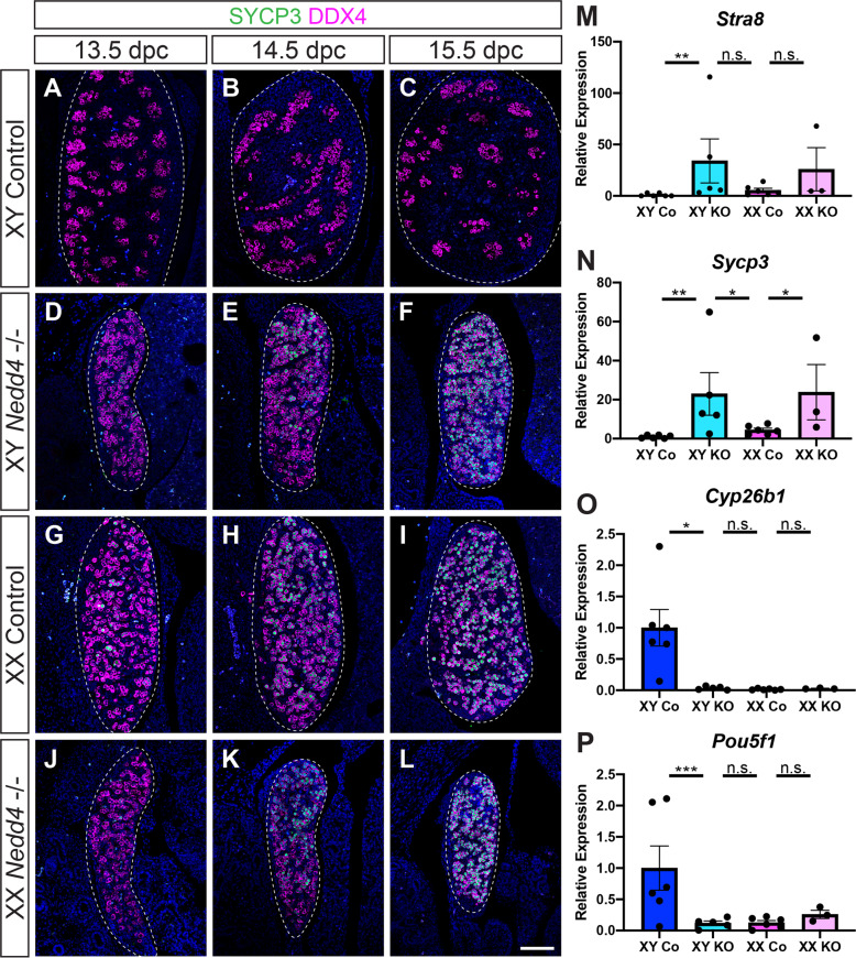
Fig. 4. Germ cells in XX Nedd4-/- mice enter meiosis.
A–L Section immunofluorescence on 13.5 dpc, 14.5 dpc and 15.5 dpc XY and XX Nedd4-/- embryos alongside XY and XX littermate controls stained for germ cell marker DDX4 (magenta) and meiosis marker SYCP3 (green). Gonads are denoted by a white dotted line. The anterior pole of each gonad is positioned at the top of each panel. Scale bars = 100 μm. RT-qPCR analyses of Stra8 (M), Sycp3 (N), Cyp26b1 (O) and Pou5f1 (P) expression at 14.5 dpc on XY Nedd4-/- gonads (XY KO) (n = 5), XX KO gonads (n = 3), XY controls (Co) (n = 6) and XX controls (n = 6). Values are expressed relative to the XY controls. Mean ± SEM; t-test; n.s. not significant, *p < 0.05, **p < 0.01, ***p < 0.001.

