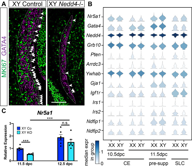
Fig. 6. Impaired proliferation and decreased Nr5a1 expression in XY Nedd4-/- mice.
A Section immunofluorescence on 11.5 dpc (18 tail somites [ts]) XY Nedd4-/- embryos and XY controls stained for gonadal somatic cell marker GATA4 (magenta) and proliferation marker MKI67 (green). The anterior pole of each gonad is positioned at the top of each panel. Gonads are denoted by a white dotted line. MKI67-positive cells within the coelomic epithelium are highlighted by white arrowheads. Scale bar = 100μm. B Violin plots showing the expression of gonadal progenitor markers Nr5a1 and Gata4 as well as Nedd4 and Nedd4 interacting proteins in mouse gonadal cells expressing both Nr5a1 and Gata4 at E10.5 and E11.5. XX and XY expression levels are shown separately. Expression levels are shown in log-normalised counts. Colour indicates median expression in the group according to the scale shown in the left bottom corner. CE coelomic epithelium, pre-supp pre-supporting cells, SLC supporting-like cells. C RT-qPCR analyses of Nr5a1 at 11.5 dpc (17–19ts, gonads + mesonephroi) and 12.5 dpc (30ts, gonads only) on XY Nedd4-/- gonads (KO) (light blue; n = 6) and XY controls (Co) (dark blue; n = 6). Values are expressed relative to 11.5 dpc XY controls. Mean ± SEM; t-test; n.s. not significant, ***p < 0.001.

