Figure 7.
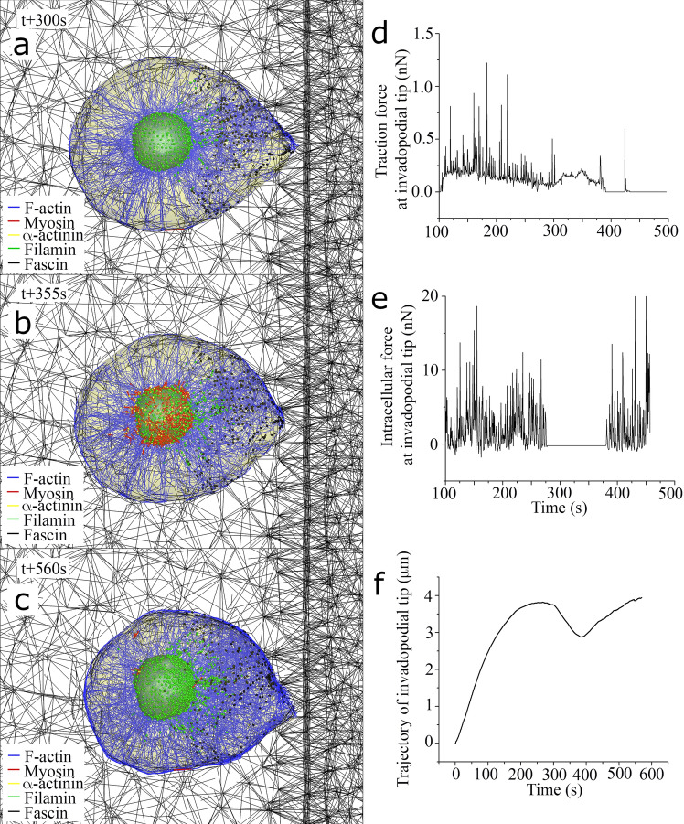
Cyclic motion of invadopodia dynamics during the directed cell migration towards stiffer ECM. Selected still shots of simulated cell migration toward stiffer ECM at time points of (a) 300 s (at the end of 1st protrusive phase), (b) 355 s (at the end of 1st retractile phase), and (c) 560 s (at the end of 2nd protrusive phase). Three graphs in (d), (e), and (f) show time-varying traction (extracellular) force, intracellular force, and trajectory at the tip of invadopodium, respectively.

