FIGURE 2.

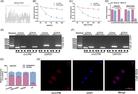
Characterization of circular CPM (circCPM). (A) Validation of head‐to‐tail splicing of circCPM using Sanger sequencing. (B and C) The relative expression changes of circCPM and CPM mRNA in AGS‐5FU and HGC‐27‐5FU after actinomycin D treatment for 4 h, 8 h, 12 h and 24 h. (D) The relative expression changes of circCPM and CPM mRNA in AGS‐5FU and HGC‐27‐5FU after RNase R treatment. (E and F) RT‐PCR‐based detection of circular and linear CPM using convergent and divergent primers in cDNA and genomic DNA (gDNA). (G) qRT‐PCR analysis confirming that circCPM and linear CPM are mainly located in the cytoplasm. (H) Fluorescence in situ hybridization (FISH) results depicting the cytoplasm location of circCPM. Scale bar = 5 μm. (Graph represents mean ± SD; *p < .05, **p < .01 and ***p < .001)
