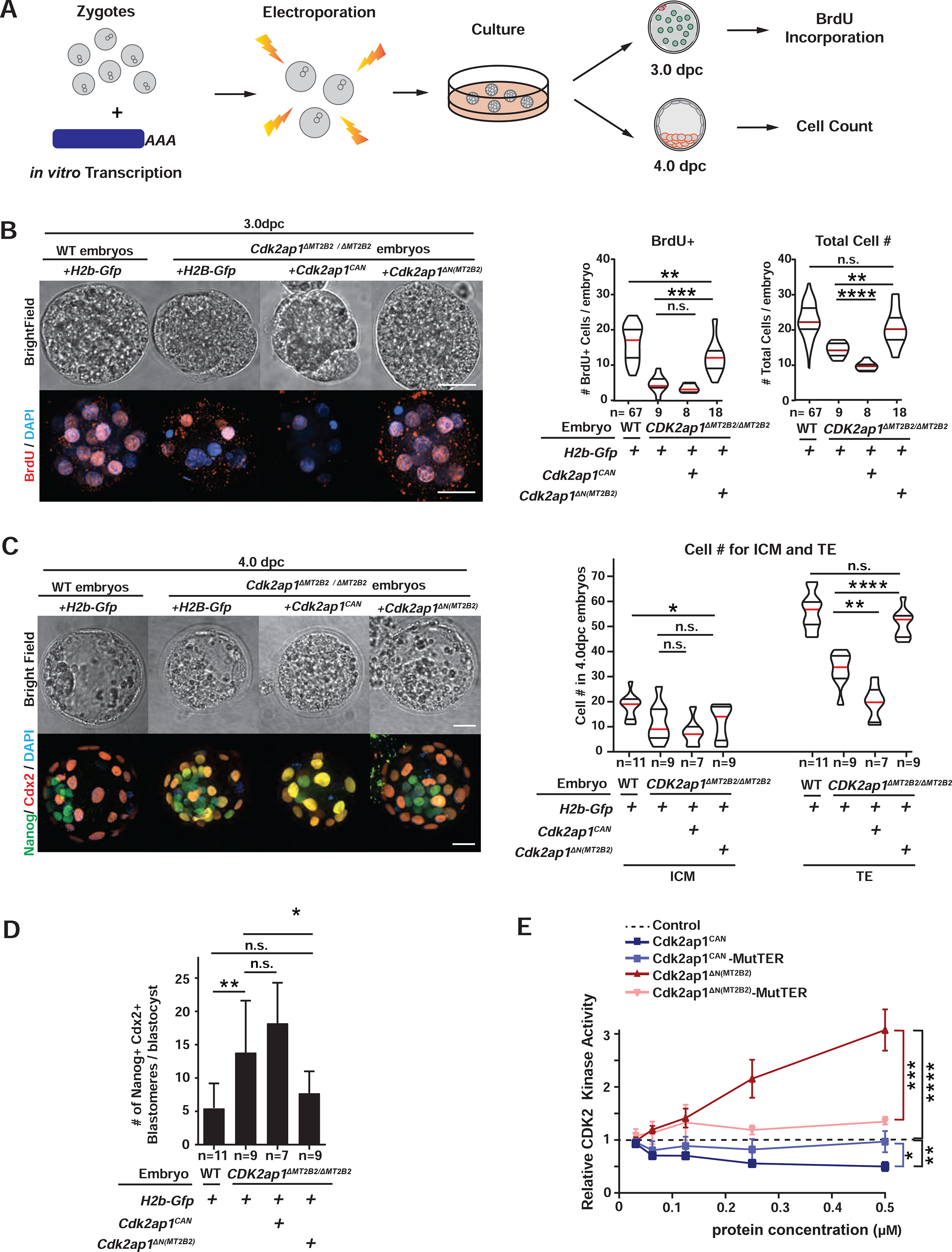
Figure 4. Cdk2ap1ΔN (MT2B2) and Cdk2ap1CAN have opposite effects in cell proliferation.

A. Diagram illustrates the experimental scheme for mRNA electroporation into zygotes. B, C. Cdk2ap1CAN and Cdk2ap1ΔN (MT2B2) have opposite effects on S-Phase entry and cell proliferation. H2b-Gfp, Cdk2ap1CAN or Cdk2ap1ΔN (MT2B2) mRNAs were each electroporated into Cdk2ap1ΔMT2B2/ΔMT2B2 zygotes, and (B) resulted morula were compared for BrdU incorporation at 3.0 dpc. Ectopic expression of Cdk2ap1ΔN (MT2B2) restores S-Phase entry and cell proliferation in Cdk2ap1ΔMT2B2/ΔMT2B2 embryos (B). Representative images (left) and quantitation of BrdU positive and total cell number (right) are shown. Violin plots are shown with median (red), as well as lower (25%) and upper (75%) quartiles (black). Scale bars, 20 μm. H2b-Gfp vs Cdk2ap1CAN in Cdk2ap1ΔMT2B2/ΔMT2B2 embryos: BrdU, n.s.; total cell number, **** P < 0.0001, t=5.7, df=15. H2b-Gfp vs Cdk2ap1ΔN (MT2B2) in Cdk2ap1ΔMT2B2/ΔMT2B2 embryos: BrdU, *** P =0.0002, t=4.5, df=25; total cell number, ** P =0.002, t=3.4, df=25. C, D. Ectopic expression of Cdk2ap1ΔN (MT2B2) rescues cell proliferation and cell fate specification defects in Cdk2ap1ΔMT2B2/ΔMT2B2 embryos. D. Representative confocal image of Cdx2 and Nanog immunostaining (left) and quantitation of ICM and TE cell number (right) are shown for 4.0 dpc Cdk2ap1ΔMT2B2/ΔMT2B2 embryos with overexpression of Cdk2ap1CAN or Cdk2ap1ΔN (MT2B2). Scale bars, 20 μm; White arrows, Nanog and Cdx2 double positive cells. H2b-Gfp vs Cdk2ap1CAN, TE, ** P =0.002, t=3.9, df=14; H2b-Gfp vs Cdk2ap1ΔN (MT2B2, TE, **** P < 0.0001, t=6.1, df=16. D. Quantitation of Nanog and Cdx2 double positive cells is shown for Cdk2ap1ΔMT2B2/ΔMT2B2 embryos overexpressing Cdk2ap1CAN or Cdk2ap1ΔN (MT2B2). H2b-Gfp-overexpressing wildtype vs. Cdk2ap1ΔMT2B2/ΔMT2B2 embryos, ** P = 0.007, t=3.1, df=17; H2b-Gfp vs Cdk2ap1ΔN (MT2B2) in Cdk2ap1ΔMT2B2/ΔMT2B2) embryos, * P =0.04, t=2.2, df=16. E. Cdk2ap1CAN and Cdk2ap1ΔN (MT2B2) have opposite effects on Cdk2 kinase activity. Recombinant Cdk2ap1CAN, Cdk2ap1CAN-MutTER, Cdk2ap1ΔN (MT2B2) or Cdk2ap1ΔN (MT2B2)-MutTER protein was incubated with recombinant CDK2, CYCLIN E, and HISTONE H1 in vitro to assay their effects on CDK2 activity at different concentrations. Three independent experiments were performed. Dashed line, baseline CDK2 kinase activity with elution buffer as the “control” input. Error bars, s.e.m. Control vs Cdk2ap1CAN, **P = 0.001, t=8.4, df=4. Cdk2ap1CAN vs Cdk2ap1CAN-MutTER, *P = 0.02, t=3.8, df=4. Control vs Cdk2ap1ΔN (MT2B2), ****P < 0.0001, t=10.5, df=6; Cdk2ap1ΔN (MT2B2) vs Cdk2ap1ΔN (MT2B2)-MutTER, ***P = 0.0003, t=8.9, df=5. All P values were calculated using unpaired, two-tailed Student’s t test. n.s., not significant. See also Figure S4.
