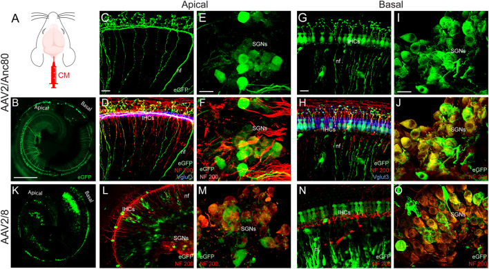
FIGURE 3.
Effects of CM injection of AAV on cochlear neural structure transduction efficiencies. (A): schematization of the CM injection technique. (B,K): Representative mosaic confocal images showing eGFP-positive cochlear structures in whole-mount preparations of cochleae 15 days after CM injection with AAV2/Anc80L65 (B) and AAV2/8 (K). Scale bar: 400 µm. (C,D,G,H,L,N): Representative magnification of organs of Corti from the apical (C,D,L) and basal regions (G,H,N) and the spiral ganglions from the apical (E,F,M) and basal regions (I,J,O) of the cochleae 15 days after CM injection of AAV2/Anc80L65 (C–J) and AAV2/8 (L–O). (C,E,G,I): eGFP (green)-positive auditory-nerve fibre terminals (nf, C,G), inner hair cells (IHCs, G), and spiral ganglion neurons (SGNs, E, I). (D,F,H,J, L–O): Merged images. NF200 immunolabelled auditory-nerve fibers and somata of the SGNs are in red. Vglut3 immunolabelled IHCs are in blue. Fy: fibrocytes. Scale bars: 20 µm.

