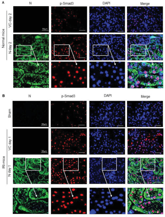
Figure 2.

Tow‐color immunofluorescence detects that SARS‐CoV‐2 N protein is highly expressed by TECs and is colocalizing with activated Smad3 (p‐Smad3) in nuclei of TECs. A) SARS‐CoV‐2 N expression and activation of Smad3 signaling in normal mice. B) SARS‐CoV‐2 N expression and activation of Smad3 signaling in IRI‐mice. Note that SARS‐CoV‐2 N protein is highly expressed by TECs in a granular pattern and is colocalized with p‐Smad3 in nuclei of TECs. DAPI (blue, nuclei), p‐Smad3 (red), SARS‐CoV‐2 N protein (green). Scale bar = 50 µm.
