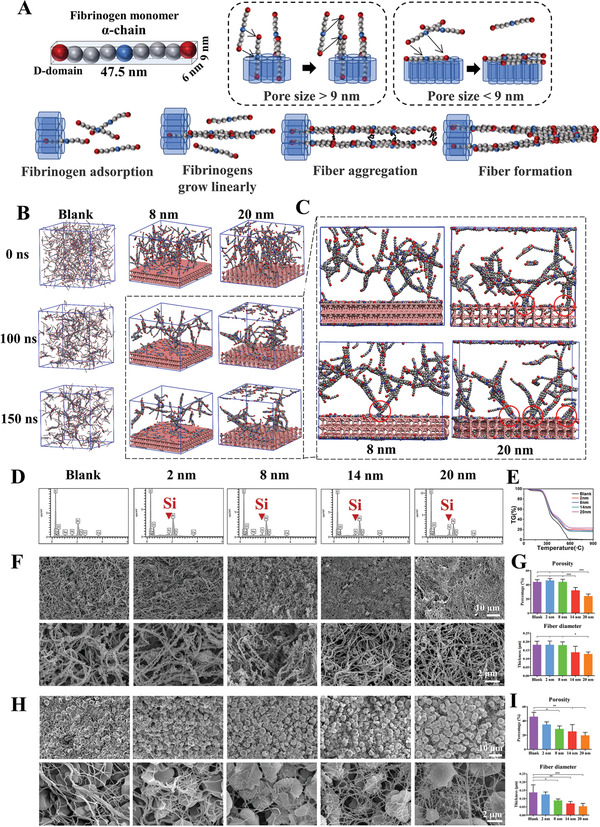
Figure 4.

The mesopore sizes above 9 nm generate thinner fibers via regulating fibrinogen behaviors. A) The model fibrinogen monomer α‐chain is presented, showing the size of fibrinogen D domain is 6 nm × 9 nm. When the mesoporous size is smaller than 9 nm, the fibrinogen D domain cannot enter the mesopore and the entire fibrinogen structure is adsorbed on the mesoporous structure surface. Opposite situation can be observed in the > 9 nm group. Part of the fibrinogen enters the pore structure, with the remaining part becoming exogenous nucleus. B) Molecular dynamics simulation diagrams of fibrinogen aggregation in the blank, 8 nm, and 20 nm groups. C) The adsorption sites of the 8 and 20 nm groups in enlarged view. The interaction sites of mesoporous silica and fibrin are labeled as red circles. D) Element distribution of mesoporous silica‐regulated fibrin network using SEM‐EDS. E) TG detection of mesoporous silica‐regulated fibrin gel. F) SEM observation of mesoporous silica‐regulated fibrin gel. G) The porosity and diameter measurement of mesoporous silica‐regulated fibrin gel. H) SEM observation of mesoporous silica‐regulated blood clot fibrin network. I) The porosity and diameter measurement of mesoporous silica‐regulated blood clot fibrin network. Data are presented as means ± s.d.; n = 3; *p < 0.05, **p < 0.01, ***p < 0.001 by one‐way ANOVA with Tukey's post hoc test.
