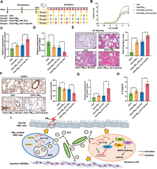
Figure 5.

EV‐packaged let‐7i‐5p induces asthma in vivo. Thirty‐two female BALB/c mice were randomly assigned to the OVA group (group 1), OVA+PM2.5 group (group 2), OVA+PM2.5+NC‐EVs group (group 3), or OVA+PM2.5+let‐7i‐5p‐EVs group (group 4). A) Schematic showing the process used to establish the mouse model. The green arrows indicate that each mouse was injected with 28.8 µL of the OVA suspension containing 3.6 µg of OVA and 144 µg of Al(OH)3. The yellow arrows indicate that mice were exposed to 2% OVA for 20 min. The grey arrows indicate that mice were exposed to 1.57 mg kg−1 PM2.5. The blue arrows indicate that mice were challenged by an endotracheal instillation of 2 µg of NC‐EVs. The red arrows indicate that mice were challenged by an endotracheal instillation of 2 µg of let‐7i‐5p‐EVs. B) Detection of bronchoconstriction using myography. C) The level of EV‐packaged let‐7i‐5p in BALF was determined using RT‐qPCR. D) The level of DUSP1 in BALF was determined using RT‐qPCR. E) Representative images of H&E staining in lung tissues and total lung injury scores for each group. Scale bar, 50 µm. F) Representative photographs and quantification of DUSP1 immunostaining in lung tissues. Scale bar, 50 µm. G) The level of let‐7i‐5p in lung tissues was determined using RT‐qPCR. H) The concentration of IL‐6 in BALF was measured using ELISA. I) In the proposed model, EV‐packaged let‐7i‐5p secreted by PM2.5‐treated HBE cells induce asthma by regulating the expression of its target gene DUSP1 and activating the MAPK signaling pathway in recipient cells. MVB, multivesicular bodies. Statistical significance was assessed using two‐tailed Student's t‐test. Values represent means ± SD. * p < 0.05.
