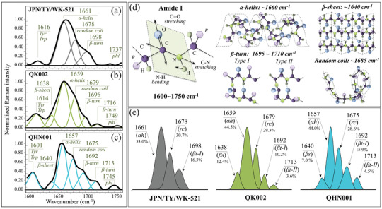
Figure 5.

Spectral Zone IV (Amide I, 1600–1750 cm−1) of the Raman spectra of a) JPN/TY/WK‐521, b) QK002, and c) QHN001 viral strains; spectra are deconvoluted into a sequence of Gaussian‐Lorentzian sub‐bands (frequencies for selected bands shown in inset). The abbreviations Trp and Tyr refer to tryptophan and tyrosine, respectively. d) Schematic drafts of the Amide I vibrational mode, the different secondary structure of proteins and related frequencies; in e), signals are shown, which are used to estimate the fractions of different protein secondary structures (shown in inset together with the frequencies of the selected signals) found in different strains. The abbreviations βs, αh, rc, and βt‐I, βt‐II, and phl represent β‐sheet, α‐helix, random coil, two types of β‐turn rotamers, and phospholipids, respectively.
