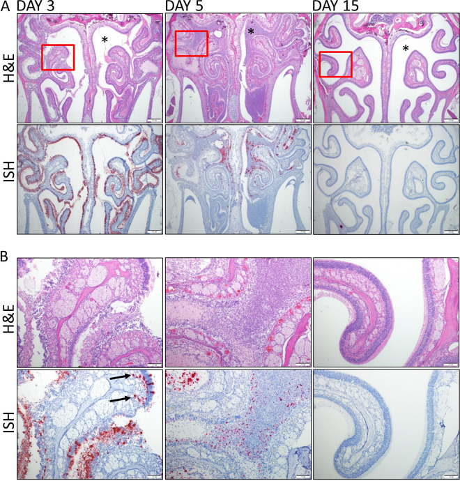
FIG 3.
SARS-CoV-2 infection of the nasal cavity. (A) Representative H&E and ISH staining of hamsters’ nasal cavities on days 3, 5, and 15. Corresponding ISH images are shown below the H&E stains, with viral labeling in red and with marked staining of the nasal and olfactory epithelium on day 3. Viral genomic RNA (red) was detected on day 3, decreased on day 5, and was absent on day 15. ISH panels were counterstained with hematoxylin (blue). Nasal cavity exudate was limited on day 3 and absent on day 15 but partially occluded the cavity on day 5 (asterisks). (B) Higher magnification of the area in the box in the top image. The day 3 ISH image shows viral labeling in the deeper lamina propria and olfactory nerve layer below the olfactory epithelium (arrows).

