FIG 4.
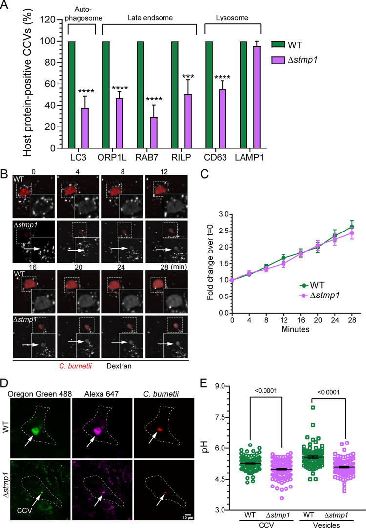
Δstmp1 CCVs preferentially fuse with lysosomes and become more acidic. (A) Quantification of CCVs positive for markers of host autophagosomes, late endosomes, and lysosomes indicates that Δstmp1 CCVs are deficient in autophagosome and late endosome markers. Infected cells were stained by immunofluorescence for CD63 or LAMP1 or transfected with LC3-GFP, ORP1L-GFP, RAB7-GFP, or RILP-GFP and analyzed by fixed microscopy. Images were visually scored for the presence or absence of the host proteins on the CCV at 3 dpi. Data are shown as the mean ± SEM of at least 20 CCVs in each of three independent experiments. Statistical significance was determined by multiple t tests; ***, P < 0.005; ****, P < 0.001. (B) Representative images of fluorescence dextran trafficking to the CCV at 3 dpi. mCherry-expressing WT- or Δstmp1 mutant-infected HeLa cells were pulsed with Alexa 488 dextran for 10 min, followed by live cell spinning disk confocal microscopy, where the cells were imaged at 0 min postpulse and then every 4 min for 28 min. (C) Quantification of changes in dextran fluorescence intensity reveal no significant difference in dextran trafficking to WT and Δstmp1 CCVs. The fluorescence intensity of Alexa 488 dextran was measured from an identical region of interest (ROI) within the CCV at each time point. The mean fold change of fluorescence intensity over the initial time point (t = 0) was plotted against time. Data are shown as the mean ± SEM of at least 20 CCVs in each of three independent experiments. Statistical significance was determined by multiple t tests. (D) The pH of CCV and host cell endosomes was determined at 3 dpi using a ratiometric fluorescence assay. mCherry-expressing WT- or Δstmp1 mutant-infected HeLa cells were labeled with Oregon green 488 and Alexa Fluor 647 dextran for 4 h followed by a 1-h chase. (E) Z-stacked images were acquired by live cell spinning disk confocal microscopy, and Oregon green 488, and Alexa 647 intensities were quantitated for each CCV and host cell endosomes and compared to a standard curve to generate individual CCV pH measurements and mean endosomal pH. Δstmp1 CCVs are significantly more acidic than WT CCVs. Increased acidification of mature endosomes in Δstmp1 mutant-infected cells indicates that Δstmp1 mutant bacteria are unable to completely block endosomal maturation at 3 dpi. Data are shown as the mean ± SEM of at least 30 cells in each of three independent experiments. Each circle represents an individual cell or CCV. Statistical significance was determined by one-way ANOVA with Tukey’s post hoc test.

