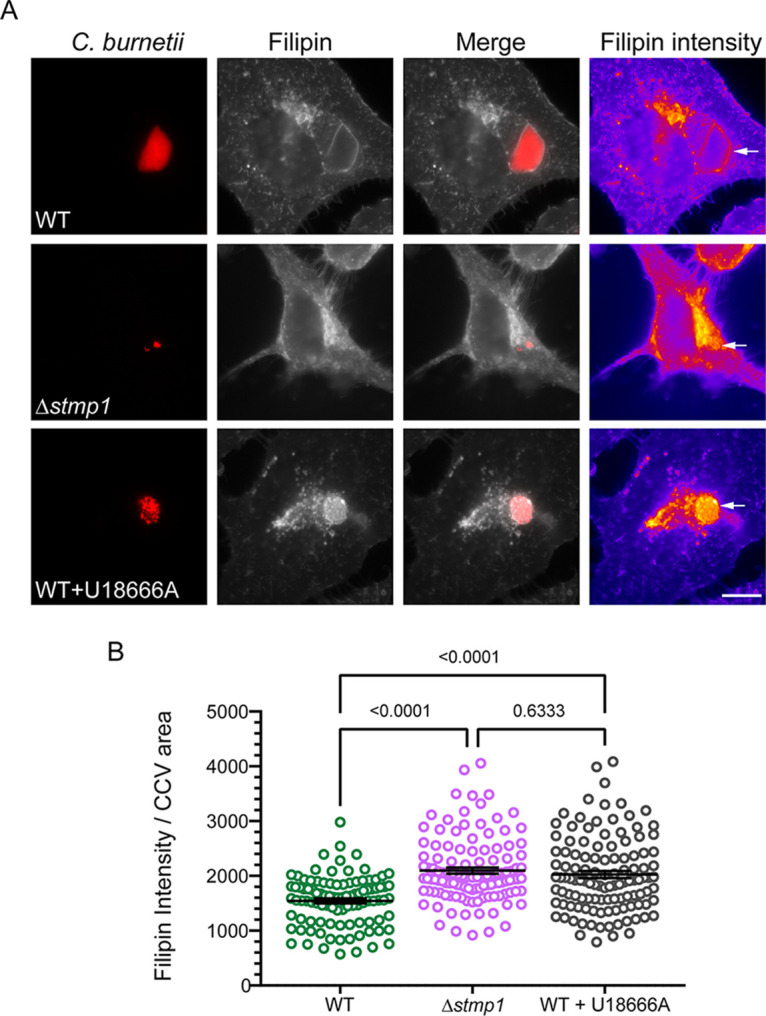
FIG 5.

The Δstmp1 mutant CCV accumulates cholesterol. Relative cholesterol levels, measured by filipin labeling, indicate that the Δstmp1 CCVs have elevated cholesterol. HeLa cells were infected with mCherry-expressing WT or Δstmp1 mutant bacteria and sterols stained at 3 dpi using filipin. As a positive control, WT-infected cells were treated for 4 h with 5 μM U18666A, a drug that traps cholesterol in lysosomes and CCVs. (A) Filipin is shown as a heat map, with yellow showing the highest filipin intensity and blue showing the lowest filipin intensity. The white arrows point to the CCVs. Bars = 10 μm. (B) Filipin intensity was measured using ImageJ and normalized to the CCV area. Each circle represents an individual CCV. Data are shown as the mean ± SEM of at least 30 CCVs per condition in each of three independent experiments. Statistical significance was determined by one-way ANOVA with Dunnett’s post hoc test.
