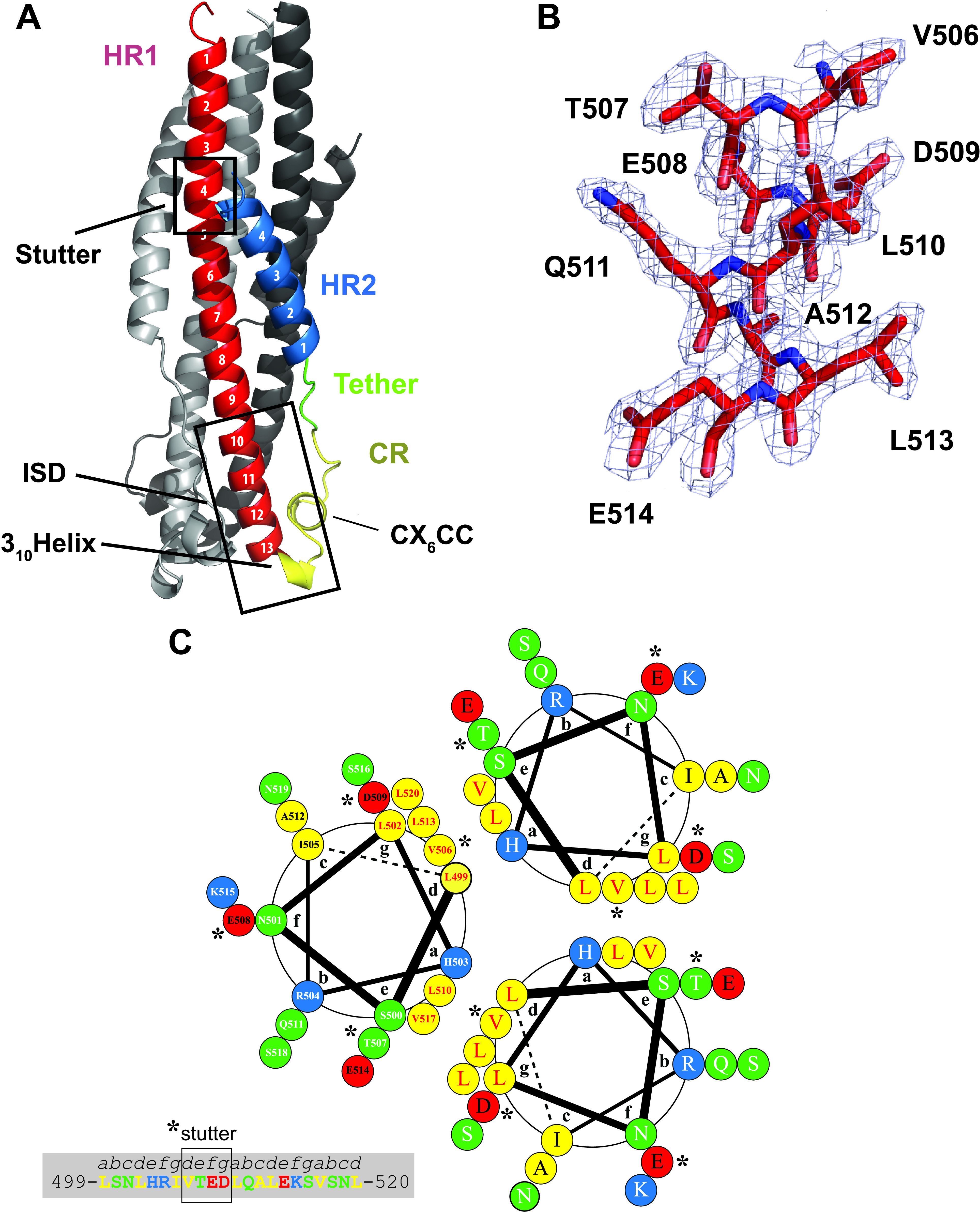
FIG 2.

PERV TM displays a 6HB stabilized by electrostatic interactions. (A) Annotated ribbon representation of the PERV TM493–587 structure. The frontmost protomer is color-coded according to the schematic presented in Fig. 1A. Key functional and structural regions of the PERV TM are annotated. (B) Representative 2|Fo| − |Fc| electron density map contoured at 1σ and superimposed on the final refined PERV TM structure. Strong, well-defined electron density is observed throughout the HR1, CR, tether, and HR2 regions. (C) Helical wheel analysis of the central PERV HR1 trimer coiled-coil from residues 499 to 520 (helix turns 2 to 6) with the heptad repeat labeled a to g. Hydrophobic, polar, negatively charged and positively charged residues are colored yellow, green, red, and blue, respectively. Residue numbering is presented in one of the helical protomers. The presence of 506VTED509 in the PERV TM (highlighted with asterisks) results in a stutter of the heptad repeat, where it disrupts the periodicity of hydrophobic residues at the a and d positions of the heptad repeat.
