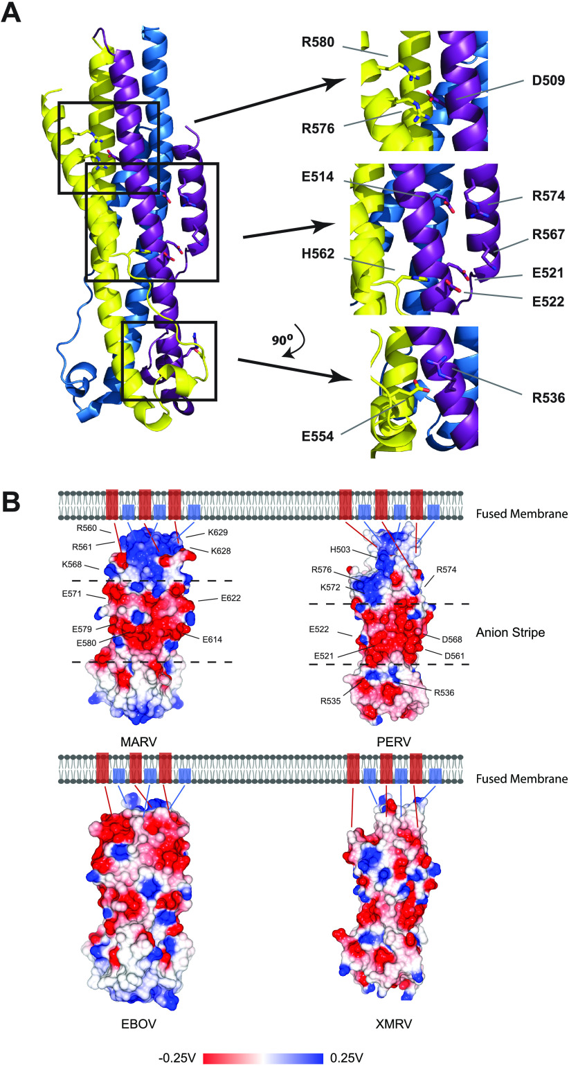
FIG 3.
PERV TM structure displays a central anion stripe. (A) Ribbon representation of PERV TM intermolecular electrostatic cross-links. Three sets of complex intermolecular salt bridges are observed between protomers R576-D509-R580, E522-H562, and E554-R536. Residues E521 and E522 that contribute to the central anion stripe are indicated. (B) Surface electrostatic potential map calculated for postfusion EBOV, MARV, PERV, and XMRV TMs. Negatively charged anion stripes are displayed along the midsection of the MARV and PERV surfaces and are found between dashed lines that intersect the surfaces. Acidic residues that contribute to the anion stripe and basic residues that line the anion stripe are annotated. Fusion proteins without anions stripes from the same viral family are found on the bottom panel. Blue and red rectangles found in the membrane correspond to the N-terminal fusion peptide and C-terminal transmembrane domain, respectively. Electrostatic potential is given in units of volts.

