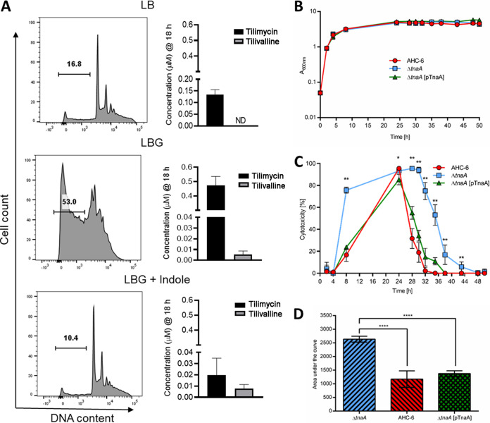
FIG 5.
Enterotoxicity of K. oxytoca complex is differentially regulated by glucose and indole. (A) Filtered supernatants from K. grimontii strain UCH-1 cultures, grown for 18 h in LB medium, LBG, or LBG plus 1.0 mM indole, were applied to T84 enterocytes. Shown are percentages of sub-G1 apoptotic cells following 72 h of exposure as determined by flow cytometry. Concentrations of tilimycin and tilivalline in the culture supernatants were determined by LC-MS (n = 3 to 8). The data are representative images or mean values ± standard errors of the means. (B) Growth comparison of indole-producing K. oxytoca AHC-6, the indole-deficient AHC-6 ΔtnaA, and complementing strain AHC-6 ΔtnaA (pTnaA) grown in CASO medium at 37°C (n = 3). The data represent mean values ± SD. (C) Cytotoxicity of filtered supernatants collected from cells used for panel B on HeLa cells (n = 3; measured in triplicates). Shown are reciprocal values (means ± SD) of surviving HeLa cells after treatment with 1/27 dilutions of supernatants. Statistical comparison (Mann-Whitney) of AHC-6 and AHC-6 ΔtnaA was done for each time point. *, P < 0.05; **, P < 0.01. (D) Statistical analysis (ANOVA, Tukey) of cytotoxicity over time. Compared are mean values (±SD) of calculated area under the curve. ****, P = 0.0001.

