FIG 1.

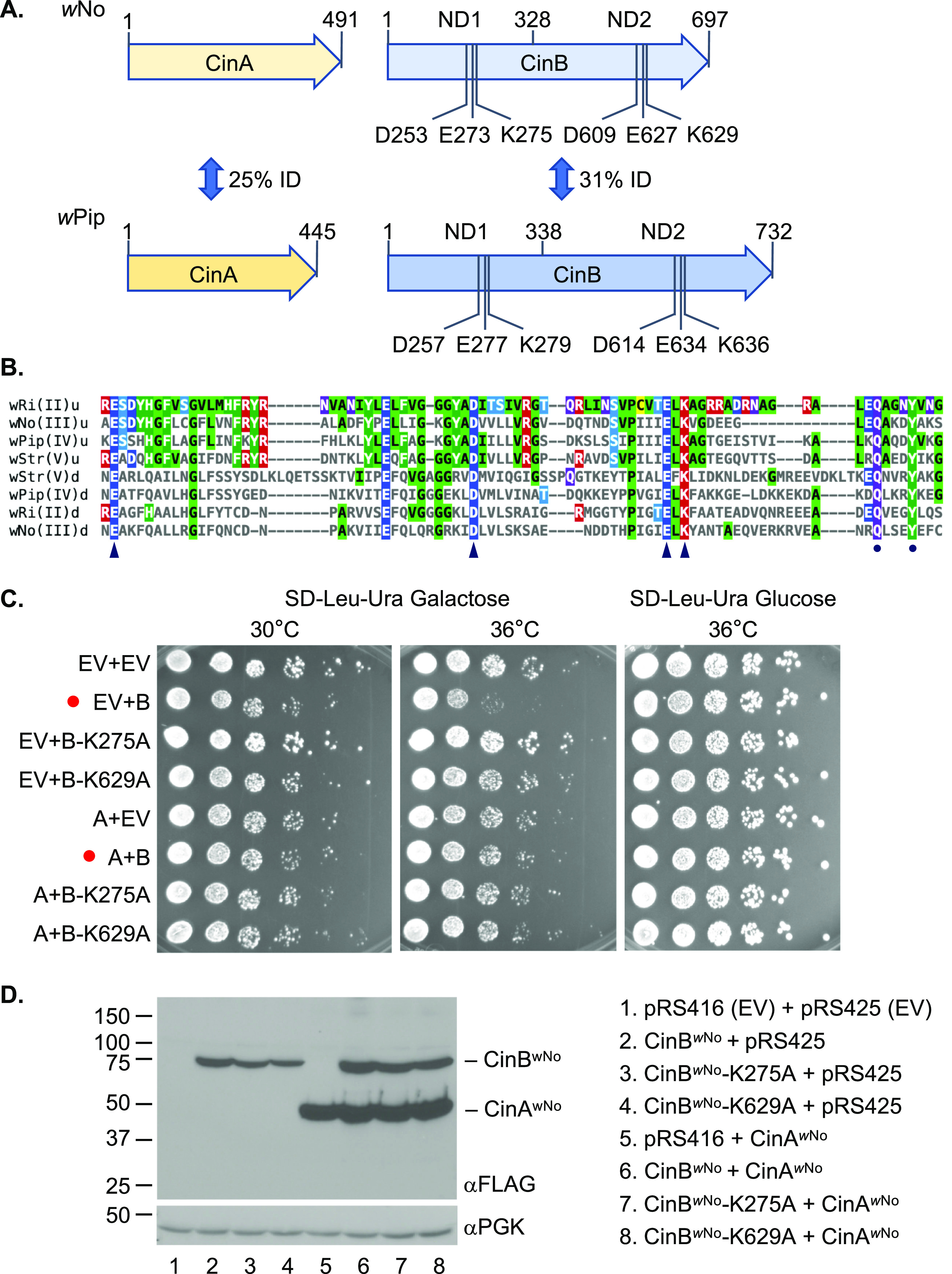
The wNo-derived CinA and CinB proteins. (A) Comparison of the wNo and wPip CinA and CinB CI factors. The wNo proteins are from the type III clade of CI factors; the wPip proteins belong to the type IV branch. Five base pairs separate the stop codon of wNo cinA from the start codon of cinB; in the wPip cinA-cinB operon, this gap is 51 bp. All CinB proteins include two intact nuclease domains (NDs), which both appear to be necessary for DNase activity and biological function. ID, identity. (B) Core sequences of the PDDEXK nuclease domains (NDs) from the four known clades of CifB factors thought to encode active nucleases (types II to V). The conserved residues that constitute the active site based on the CinBwPip (type IV) crystal structures (24) are indicated by arrowheads. A conserved QxxxY motif just downstream of the active site residues is marked by dots; this motif is characteristic of RecB-like nucleases and has been suggested to function in DNA binding (33). Alignments were done with Clustal Omega, and the figure was made using MView (1.63). u, upstream or N-terminal ND; d, downstream or C-terminal ND. (C) Growth assays of yeast expressing wNo cinA and cinB alleles. BY4741 cells were transformed with plasmids expressing the indicated alleles from a galactose-inducible GAL1 promoter, and cultures were spotted in serial dilution onto selection plates with the indicated carbon source and grown for 2.5 days at either 30°C or 36°C. Red dots highlight strains expressing WT CinBwNo with and without CinAwNo coexpression. EV, empty vector; A, CinAwNo; B, CinBwNo. (D) Expression levels of different CinA and CinB proteins in the same BY4741 transformants from panel C were measured by immunoblot analysis. Both proteins were tagged with a FLAG epitope. The anti-PGK immunoblot served as a loading control.
