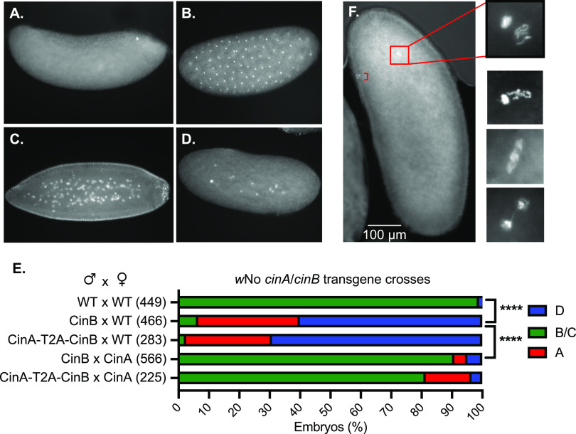
FIG 4.
CI-like embryonic defects caused by expression of CinBwNo or CinA-CinBwNo in males. (A to D) Representative images of early embryos with DNA staining by propidium iodide. (A) Unfertilized or very early arrest embryo; (B) normal embryo at ∼1 h of development; (C) normal embryo at ∼2 h of development; (D) embryo with early mitotic failure. (E) Quantification of embryo cytology into classes A to D. Embryos exhibiting normal cytology at 1 to 2 h were grouped together and are shown in green. ****, P < 0.0001 by χ2 test comparing normal (B and C) and abnormal (A and D) cytological phenotypes. All transgenic strains were confirmed by PCR (12), and all strains used in the test crosses carried the MTD-GAL4 driver except for the CinA-T2A-CinBwNo strain. The number of embryos examined for each cross is in parentheses. (F) Images of Hoechst 33342-stained embryos from transgenic CI crosses between transgenic CinBwNo males and (uninfected) wild-type females showing apposed but asynchronous male and female pronuclei with defects at the first zygotic mitosis. Embryos were fixed after allowing 30 min for egg laying. Bracket in the low-magnification image marks the three polar bodies.

