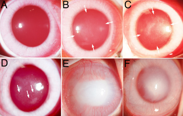
Figure 1.

Broad beam slit lamp photos of corneas after injuries that produce opacity. (A) Normal unwounded rabbit cornea with high transparency such that only the red reflex is seen for the most part. (B) Rabbit cornea one month after −3 D PRK with mild opacity (haze) in the area of the excimer laser ablation between the arrows. (C) Rabbit cornea one month after −9 D PRK with moderate opacity (haze) owing to the development of myofibroblasts and fibrosis in the area of the excimer laser ablation between the arrows. (D) Rabbit cornea at six weeks after −9 D PRK with clear areas (arrows) called lacunae developing within the scarring fibrosis. (E) Rabbit cornea at two weeks after 5 mm central 1 M sodium hydroxide exposure for 1 minute with severe fibrosis and CNV. (F) Rabbit cornea at 4 months after 8 mm Descemetorhexis removal of the central endothelium and Descemet's membrane with persistent stromal scarring fibrosis and CNV, but a decrease since the one-month time point (not shown but see Ref. 25). Original magnification ×20.
