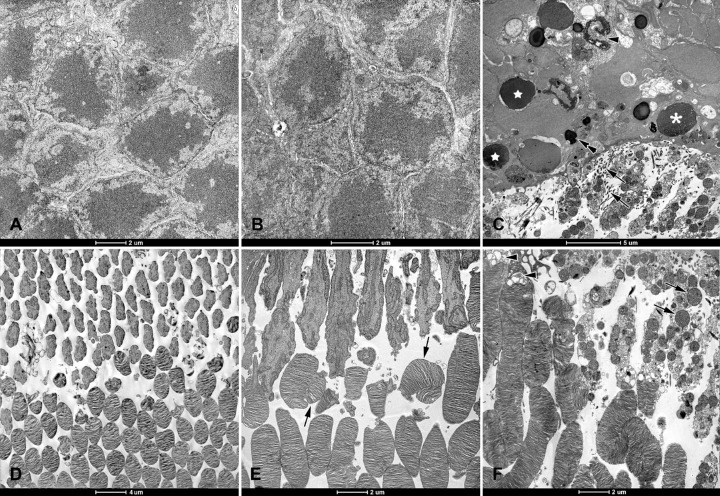
Figure 5.
Electron micrographs of retinas from Brown Norway rats. The rats were exposed to 12 hours of white light (1000 lx) and 12 hours of darkness for 10 days (control, 6 rats), 12 hours of blue light (1000 lx) and 12 hours of darkness for 10 days (long-term, 9 rats), and blue light (1000 lx) for 2 days (acute, 9 rats). Normal looking outer nuclear layers (ONL) in (A) control and (B) long term exposure groups. (C) ONL from acute exposure group. Note: Nuclei are sparser, stages of apoptosis are evident (star = pyknosis; asterisk = chromatin pyknosis and vacuolization; and arrowhead = collapsed nuclei), and multiple processes of Müller cells are present between inner segments (arrows). Pigment granules are present between scattered nuclei (double arrow). Normal looking photoreceptor outer and inner segments from (D) control and (E) long-term exposure groups. Note: Some outer segments are disarranged (arrows). (F) Inner segments from acute exposure group have disrupted membranes and swollen mitochondria (arrows). Note: Vacuolization of the transition zone between photoreceptor inner and outer segments (arrowheads). Ultrathin sections were visualized using a Tecnai 12 Spirit G2 BioTwin transmission electron microscope (FEI, USA) equipped with two digital cameras: a Veleta (Olympus, Japan) and an Eage 4k (FEI, USA).

