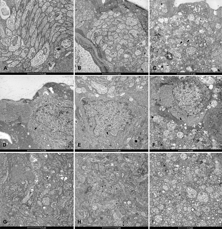
Figure 6.
Electron micrographs of retinas from Brown Norway rats. The rats were exposed to 12 hours of white light (1000 lx) and 12 hours of darkness for 10 days (control, 6 rats), 12 hours of blue light (1000 lx) and 12 hours of darkness for 10 days (long-term, 9 rats), and blue light (1000 lx) for 2 days (acute, 9 rats). In the (A) control and (B) long-term exposure groups, nerve-fiber-layer axons and their mitochondria appear normal. (C) In the acute exposure group, axons are swollen and have mitochondria with disrupted cristae (arrowheads). Somas of ganglion cells from the (D) control and (E) long-term exposure groups display normal ultrastructure with numerous free ribosomes and cisterns of rough endoplasmic reticulum (arrows). (F) Ganglion cell somas from the acute exposure group have fewer free ribosomes and cisterns of the rough endoplasmic reticulum, as well as swollen mitochondria (arrow). In retinas from the (G) control and (H) long-term exposure groups, the inner plexiform layer appears normal. (I) In the retina from the acute exposure group, the inner plexiform layer is vacuolized. Ultrathin sections were visualized using a Tecnai 12 Spirit G2 BioTwin transmission electron microscope (FEI, USA) equipped with two digital cameras: a Veleta (Olympus, Japan) and an Eage 4k (FEI, USA).

