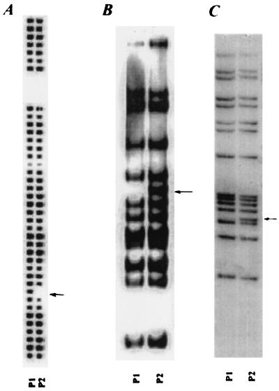
FIG. 1.
Molecular typing of the strains. (A) Spoligotype image of isolates from patient no. 1 (P1) and patient no. 2 (P2). The arrow indicates spacer 9, which was missing in the P2 isolate. (B) IS6110 RFLP fragment digested with PvuII. An arrow indicates the additional 2.5-kb band in the P2 isolate. (C) IS6110 RFLP fragment digested with SacI. The additional band is indicated by the arrow.

