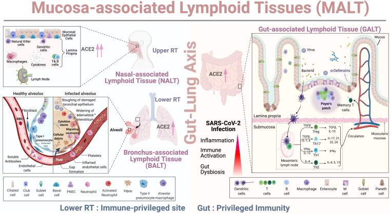
Figure 3.
Role of Mucosal and Gut Immunity in SARS-CoV-2 Infection: As shown in the schematic representation, the upper respiratory tract (URT) have specialized lymphoid structure called Nasal Associated Lymphoid Tissue (NALT). The mucosal epithelial cells covering NALT express high levels of ACE2 receptors which facilitates binding of spike protein of SARS-CoV-2. The lamina propria consisted of a mixed population of T cells, B cells, NK cells, macrophages and dendritic cells while the lower respiratory tract (LRT) is possibly an immune-privileged site as a single layer of pneumocytes forms the healthy alveolus and has very few immune cells in the vicinity. Type 1 and type II pneumocytes express ACE2 receptors and SARS-CoV-2 infection damages the respiratory epithelium, widening the interstitium followed by accumulation of fluid in the alveoli along with cellular debris. Immune cells such as neutrophils, macrophages migrate from blood vessels to infected alveolus and leads to hyperinflammation/cytokine storm, thrombosis along with disruption of the “Gut-lung axis.” The gut associated lymphoid tissue (GALT) consists of multi-follicular Peyer’s patches, plasma cells, T cells present in the lamina propria, and mesenteric lymph nodes. Dendritic cells capture microbial antigen and carry it through lamina propria, submucosa to draining mesenteric lymph node where they interact with helper T cells (Th cells). Th cells differentiate into Th1, Th2, regulatory T cells (Tregs), Th17 cells and memory T cell pool which then migrate to the gut and respond against SARS-CoV-2 infection.

