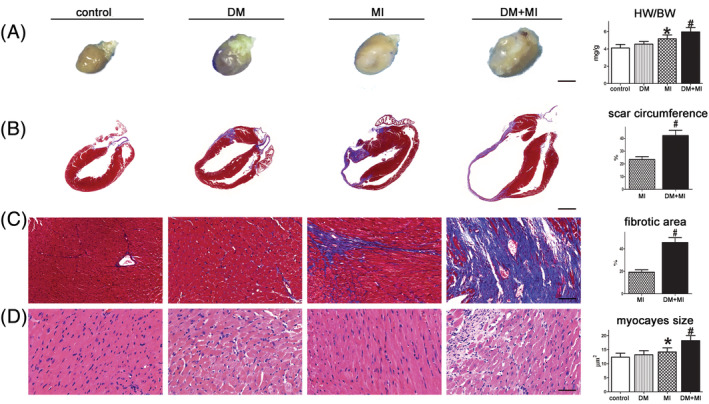
Figure 2.

DM exacerbated cardiac remodelling and myocardial injury after MI. (A) Hearts from DM + MI mice appear more spherical than those from MI and DM mice (scale bar = 3 mm). (B, C) Paraffin‐embedded sections of myocardium were stained with Masson's trichrome showing the effects of DM on scar circumference (scale bar = 5 mm) and ventricular fibrosis (scale bar = 50 μm). (D) Representative image of haematoxylin and eosin showing cardiomyocyte size from mice (scale bar = 50 μm). # P < 0.05 DM + MI vs. MI; *P < 0.05 MI vs. controls. BW, body weight; DM, diabetes mellitus; HW, heart weight; MI, myocardial infarction.
