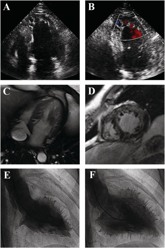
Figure 1.

Imaging of NCCM. Echocardiographic apical four‐chamber view (Panel A) showing a dilated left ventricle with trabeculations in the apical and lateral region, and blood flow in deep recesses at colour Doppler (Panel B). Cardiac MRI four‐chamber (Panel C) and axial (Panel D) view showing NCCM at lateral and apical wall. Left ventriculogram in right long‐axis oblique view during systole (Panel E) and diastole (Panel F), showing an extensive non‐compacted layer containing numerous trabeculations.
