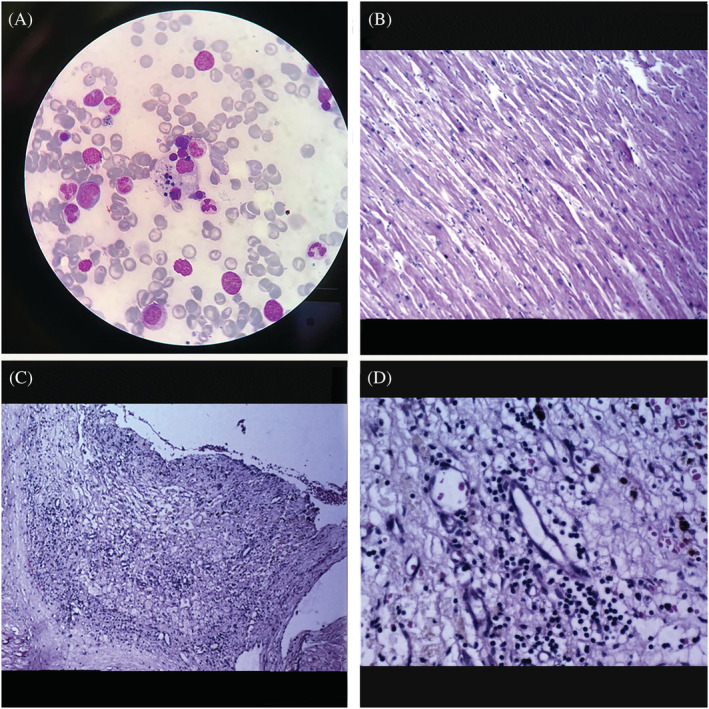
Figure 2.

(A) Bone marrow aspirates showing erythrocytes and lymphocytes engulfed by bizarrely shaped macrophage. (B) Microscopic examination shows cardiac myocytes with mild to moderate hypertrophy and delicate interstitial fibrosis. (C and D) Focus of mural granulation tissue formation and scattered chronic inflammatory cells—lymphocytic infiltration restricted to endocardium—is seen.
