Fig. 3.
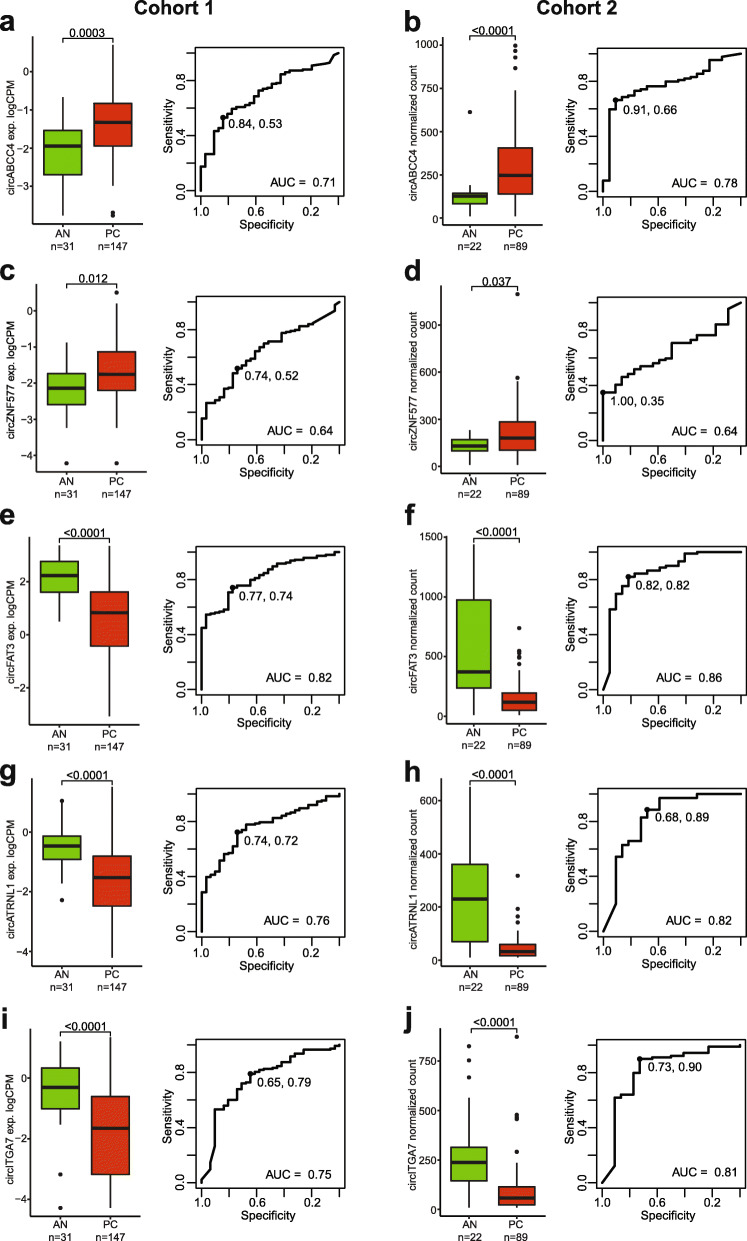
Individual circRNAs with diagnostic potential in prostate cancer. Boxplot (left) of individual circRNA expression across AN and cancer (LPC and MPC) patient tissue samples (a, c, e, g, i: cohort 1; b, d, f, h, j: cohort 2). P values represent Wilcoxon rank-sum test. Boxes represent the 25th and 75th percentiles and median. Outlier cases, defined as more than 1.5 times the IQR from the median are marked as individual dots outside the whiskers. ROC curve analysis (right) for distinguishing PC from AN tissue specimens. Specificity and sensitivity at optimal cut-off are shown

