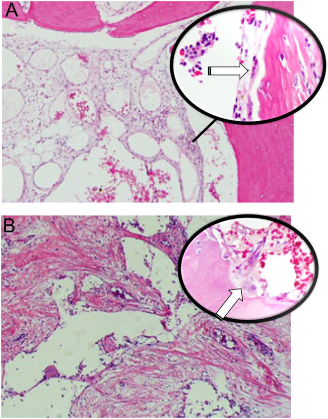
Figure 4.

Gorham–Stout disease. (A) Enlarged abnormal lymphatic channels vary in size with numerous osteoclasts (white arrow in higher magnification), (B) dilated vessels with thin walls, loss of cortex and bone trabeculae surrounded by fibrous tissue. Active osteoclasts (white arrow) are present.

 This work is licensed under a