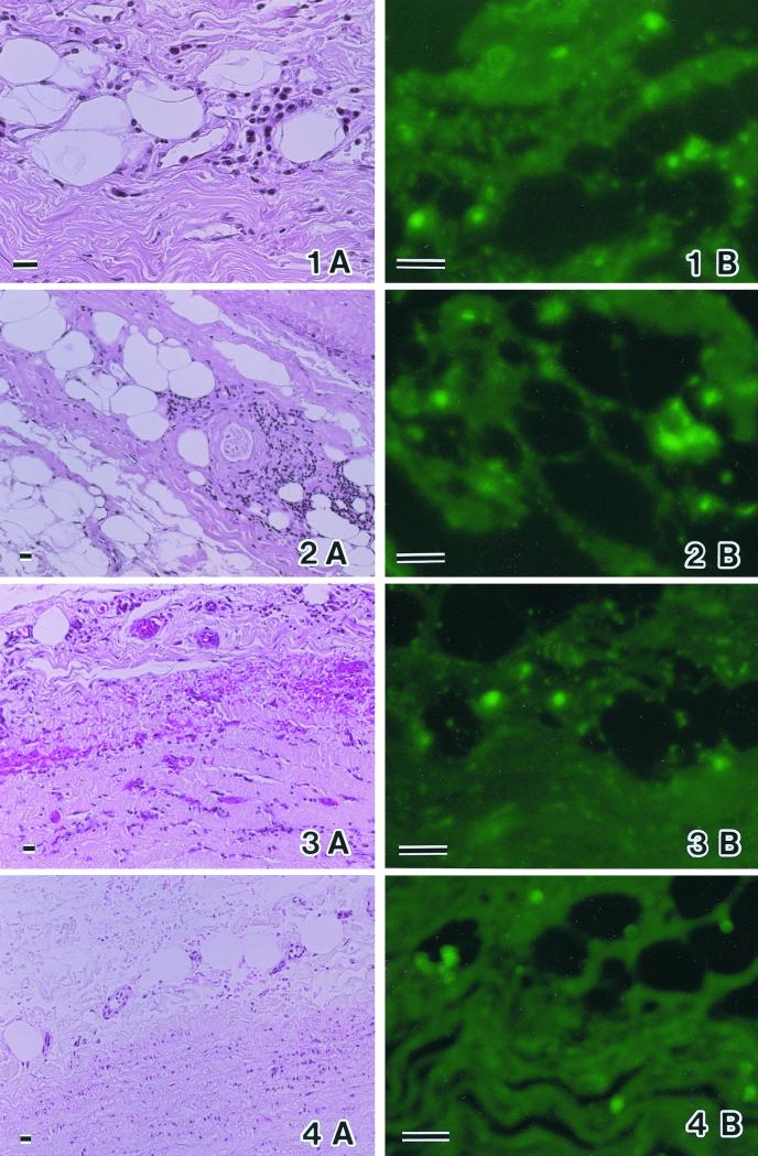
FIG. 2.
(1A, 2A, 3A, and 4A) Thin sections of atherosclerotic lesions, stained with hematoxylin and eosin, in which strong T. denticola DNA bands were detected (Table 1 and Fig. 1). (1B, 2B, 3B, and 4B) Thin sections corresponding to panels 1A through 4A, respectively, stained with rabbit antiserum against T. denticola. Clear immunofluorescent particles can be observed in localized areas of foam cells or between small muscles in panels 1B through 4B. Bars, 20 μm.

