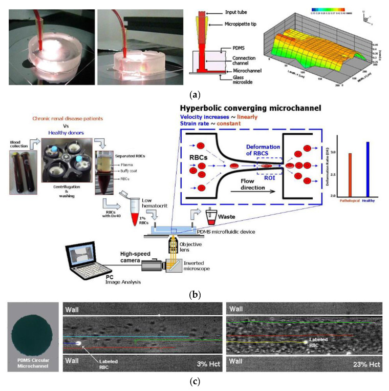
Figure 2.
Example of PDMS biomodels for hemodynamic studies: (a) rectangular PDMS microchannel to study in vitro blood and ensemble velocity profiles (U) obtained in the middle plane by means of a confocal micro-PIV system (adapted from [126]); (b) schematic diagram of the blood collection and cells deformability tests in PDMS microfluidic device (from [143]); (c) circular PDMS microchannels to study in vitro blood behavior (adapted from [152]).

