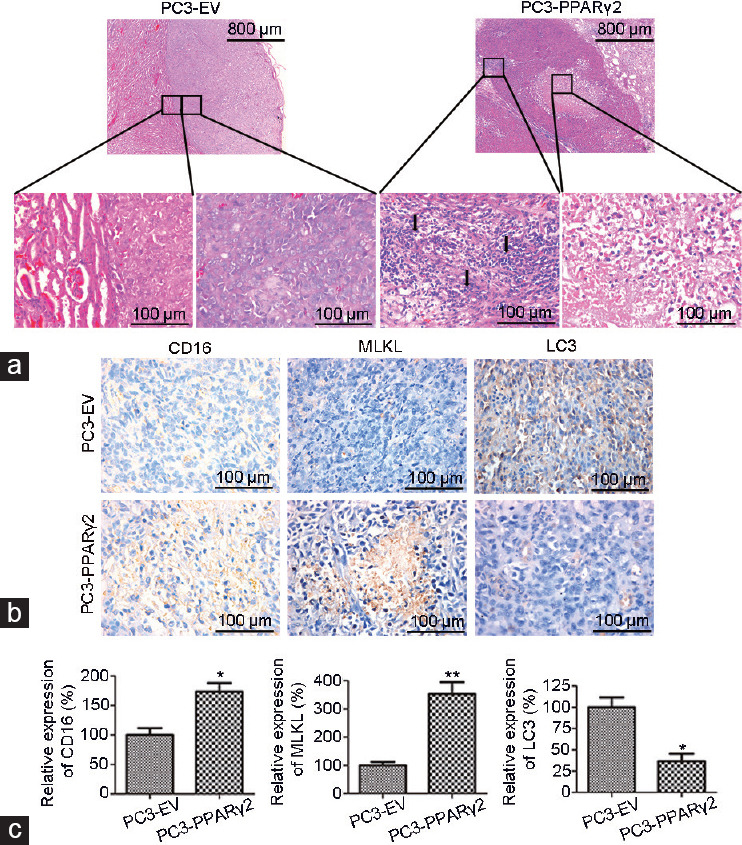
Figure 2.

Restoration of PPARγ2 isoform activity in PC3 cells induced necrosis and leukocyte infiltration in vivo. (a) Histological analyses of human prostate cancer tissue recombinants indicate significant morphological differences between the PC3-EV and PC3-PPARγ2 groups at 3 months postgrafting. PC3-EV tissue recombinants showed no leukocyte accumulation in the stroma or solid tumors (left). PC3-PPARγ2 tissue recombinants presented stromal leukocyte infiltration and necrosis of cancerous areas (right). The arrow shows leukocyte infiltration. (b) IHC staining shows low protein expression of CD16 and MLKL and high protein expression of LC3 in cancerous regions of PC3-EV tissue recombinants and high CD16 and MLKL protein expression and low LC3 protein expression in PC3-PPARγ2 tissue recombinants. (c) The relative expression of CD16, MLKL, and LC3. *P < 0.05, **P < 0.01; n = 5 for each group. PPARγ2: peroxisome proliferators-activated receptors γ 2; EV: empty vector; MLKL: mixed lineage kinase domain-like; LC3: lower microtubule-associated protein 1 light chain 3; IHC: immunohistochemistry.
