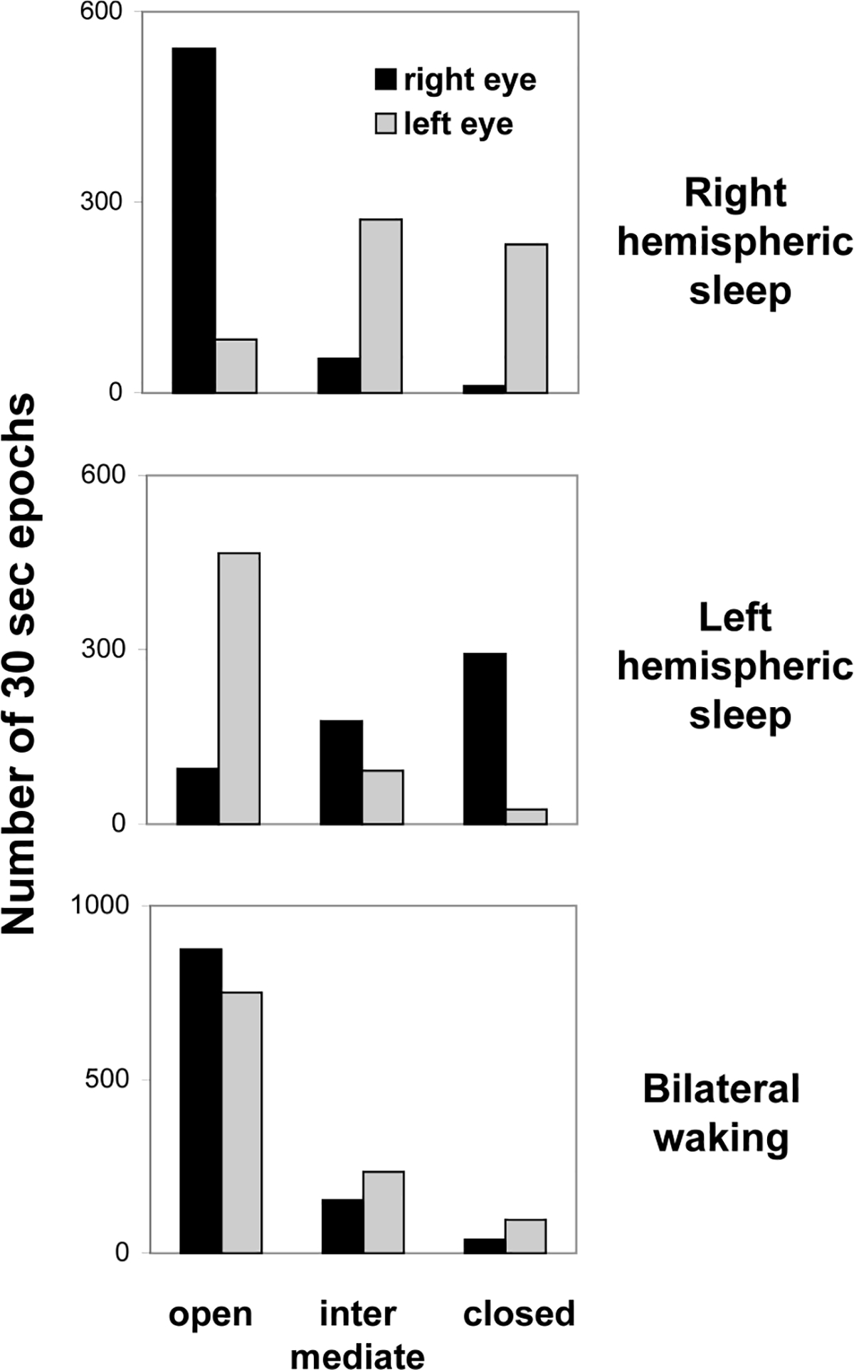
Fig. 2.

The state of the eyes during waking and sleep in a white whale. The EEG stage in each hemisphere was scored visually in 30 s. The state of the eyes was sampled in 5-s epochs and then extrapolated for consecutive 30-s epochs. Right and left hemispheric sleep represents unihemispheric SWS or asymmetrical bilateral SWS with a higher voltage EEG in the corresponding hemisphere. Reported values are the numbers of 30-s epochs with a given state of the eyes documented over 2 consecutive days.
