FIG 1.
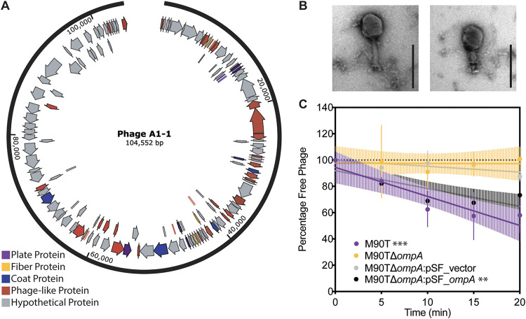
Basic characterization of newly discovered lytic phage A1-1 on host, Shigella flexneri. (A) Genome annotation of phage A1-1 using PHASTER. Plate proteins are shown in purple, fiber proteins in yellow, coat proteins in blue, phage-like proteins in red and hypothetical proteins in gray. (B) TEM image of phage A1-1 (200-nm scale bar). (C) Adsorption assay of phage A1-1 on bacterial hosts: M90T (purple), M90TΔompA (yellow), M90TΔompA:pSF_vector (gray), and M90TΔompA:pSF_ompA (black). Error bars show standard deviations of the mean for three biological replicates. Significance determined by testing whether slope of the linear regression line deviated from zero. (*, P < 0.05; **, P < 0.01; ***, P < 0.001).

