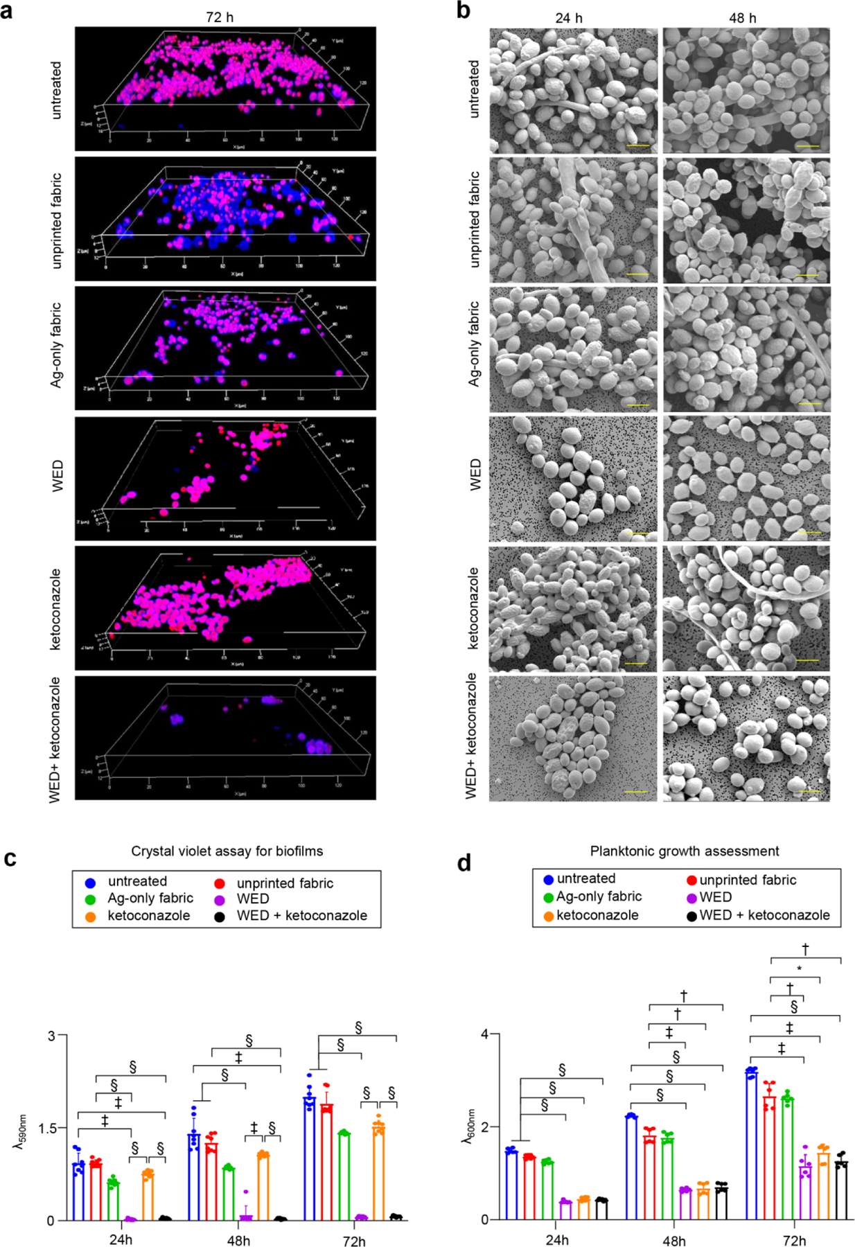
Figure 2: WED inhibited biofilm formation and planktonic growth in Candida albicans.

(a) Representative CLSM images for C albicans biofilm thickness. Biofilms were allowed to form in presence or absence of fabrics (alone or in combination with ketoconazole). After 72h, biofilms were stained with Calcofluor white and Sypro Ruby (biofilm matrix stain) and observed at 63X magnification. Display settings for all images were kept same. (b) Representative SEM images for C albicans biofilm architecture. In vitro biofilms (24h and 48h old) formed on polycarbonate membrane discs were processed for SEM and images were captured at 4000X magnification. Scale bar represents 5 µm. (c) Effect on in vitro biofilm attachment. C albicans cells were allowed to form biofilms on six well polystyrene plates, in presence of fabrics or ketoconazole. At respective time intervals, planktonic cells were washed and biofilms attached to the wells were stained with crystal violet followed by ethanol extraction and spectrophotometric analysis at 590 nm. n = 8. §P < 0.0001, ‡P < 0.0005 and *P < 0.05 (Two-way ANOVA followed by post-hoc Sidak multiple comparison test). Data are represented as the mean ± SD. (d) Effect on planktonic growth measured through absorbance. C albicans cells (untreated or with respective treatments) were cultured in YPD broth. At respective time intervals, absorbance was measured at 600 nm to assess the rate of planktonic growth. n = 6. §P < 0.0001, ‡P < 0.0005 and *P < 0.05 (Two-way ANOVA followed by post-hoc Sidak multiple comparison test). Data are represented as the mean ± SD.
