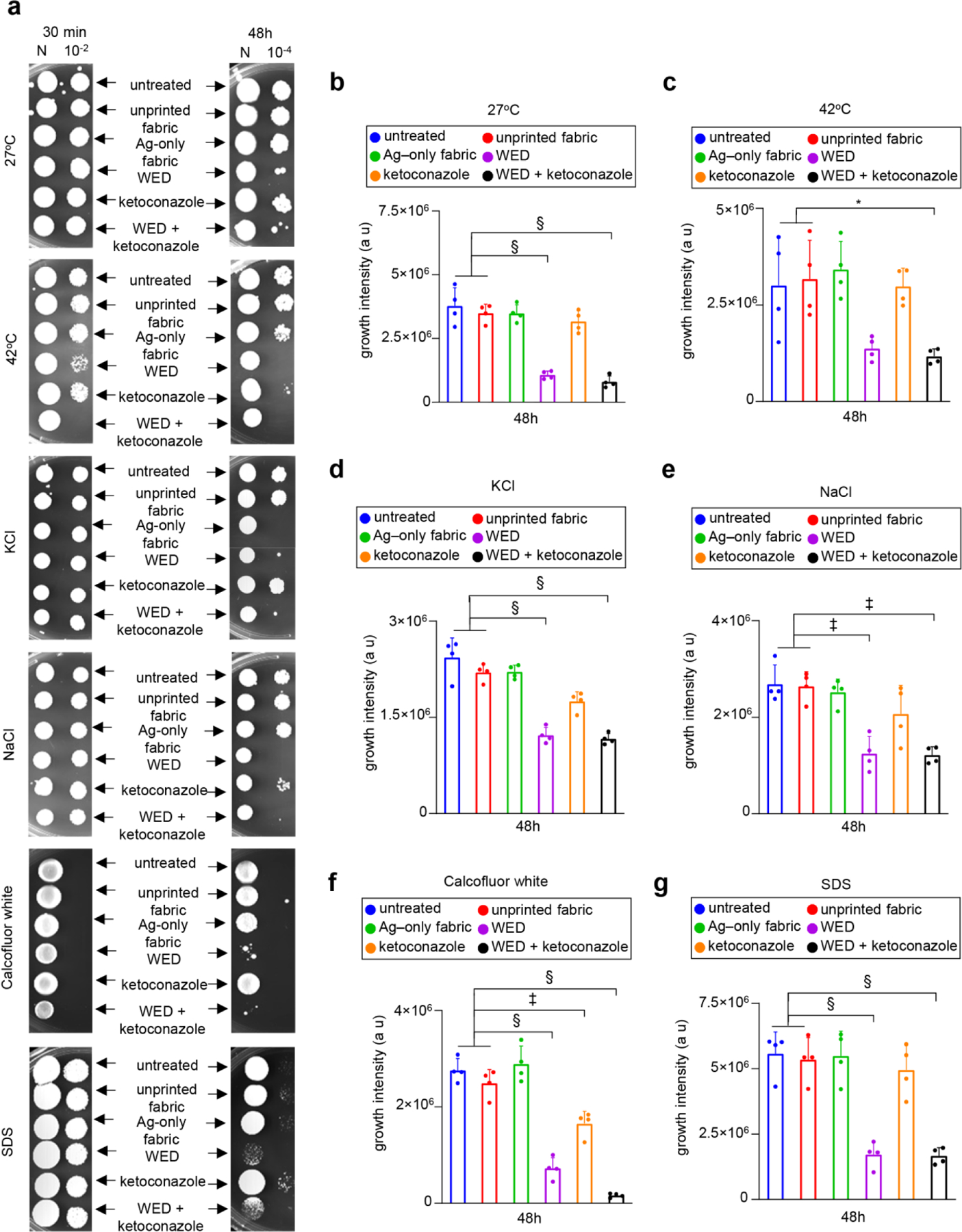
Figure 8: WED mediated Candida albicans cell wall alterations increased cell susceptibility towards secondary stressors.

(a) Representative plate images for spot viability assay with secondary cell wall stressors. Candida albicans cells were first cultured in YPD broth with fabrics or ketoconazole or a combination of both. After respective time intervals (30 min and 48h), these stressed cells were washed and spotted on to YPD agar plates with a secondary cell wall stress agent such as 1M KCl, 1M NaCl, 50 µg /ml Calcofluor white or 0.01% SDS. One set of cells were also incubated at 42°C (heat stress). All plates except heat stress plates were incubated at 27°C. After 48–72h incubation, plates were observed for growth. (b) – (g) Growth intensity measurement plots. An area of interest was selected around the growth spots on secondary cell wall stress assay plate images (after 48h) and the intensity of growth was calculated using ImageJ software. This data was graphically represented for 27°C, 42°C, KCl, NaCl, Calcofluor white and SDS respectively. n = 4 plates. §P < 0.0001, ‡P < 0.0005 and *P < 0.05 (One-way ANOVA followed by post-hoc Sidak multiple comparison test). Data are represented as the mean ± SD.
