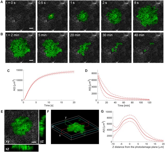
Figure 1.
Intravital multiphoton microscopy of Ca2+ waves elicited by focal photodamage in the epidermal basal layer of earlobe skin in live anesthetized mice. (A, B) Representative sequences of GCaMP6s fluorescence images acquired at shown time points from the instant preceding the beginning of the photodamage (t = 0 s) showing (A) Ca2+ wave expansion from the photodamage site (yellow asterisk in the 0.5 s image) and (B) subsequent wave waning. (C, D) Invaded area vs. time, A(t), during Ca2+ wave expansion (C) and waning (D). (C) Mean (solid line) ± s.e.m. (dashed lines) of n = 60 experiments in 14 mice. (D) mean (solid line) ± s.e.m. (dashed lines) of n = 3 experiments in 1 mouse. (E, F) Volume of tissue invaded by the Ca2+ wave after focal photodamage. (E) Views of the wave in the xy plane (focal plane) and two orthogonal (xz and yz) planes. (F) 3D rendering of the volume invaded by the wave; cyan, red and green lines indicate the xy, xz and yz planes of panel (E), respectively. (G) Invaded area vs. axial coordinate (z, along the optical axis of the objective lens); data are mean (solid line) ± s.e.m. (dashed lines) of n = 7 experiments in 1 mouse. Scale bars in A, B, E: 25 µm.

