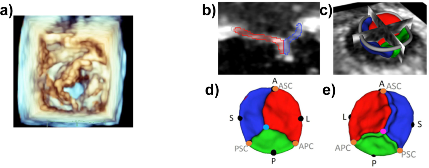
Fig.1:

A. Volume rendering of 3DE (left ventricular view) of TV in patient with HLHS; B and C. Segmentation of TV; D and E. Atrial and ventricular views of valve model with landmark annotations(A = anterior, P = posterior, S = septal, L = lateral, ASC, APC, PSC = commisures)
