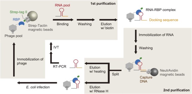
Figure 1.
Schematic illustration of PD-SELEX. RBPs are displayed on T7 phage and immobilized on Strep-Tactin magnetic beads via Strep-tag II fused to the C-terminus of RBP. After capturing RBP-binding RNAs, the phage particles are eluted, and phage-displayed RBP–RNA complexes are captured by magnetic beads immobilized with an oligo DNA complementary to the fixed RNA sequence (docking sequence). RNA and phage particles are separately eluted and amplified for the subsequent round of selection.

