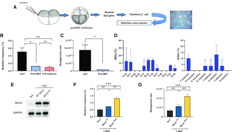
Figure 1.
Pre-MBT Xenopus embryos accumulate polymorphisms and deletions. (A) Drawing of the experimental strategy adopted to analyze mutagenesis in X. laevis embryos. Two-cell-stage embryos are injected with a supercoiled plasmid containing lacZ reporter gene (pEL1) and allowed to replicate for further three divisions. After embryo collection, plasmid DNA is extracted and transformed in lacZ-deficient bacteria for white/blue screening. (B) Mutation frequency expressed as a percentage of white colonies in each condition. The mutation frequency of lacZ recovered from embryos injected with a post-MBT amount of plasmid DNA is also included as comparison. Pre-MBT and post-MBT, n = 3; pre-injection, n = 2. (C) Mutagenesis rate in the indicated different experimental conditions expressed as mutations per base pair/locus per generation (see the ‘Materials and Methods’ section), normalized to the pre-injection background values (n = 3). (D) Mutation spectra of the lacZ gene recovered from Xenopus pre-MBT embryos after Sanger sequencing (n = 3). (E) Western blot of total protein extracts obtained from Xenopus embryos subjected to the indicated experimental conditions (n = 2). (F) Mutation frequency and (G) mutagenesis rate of lacZ isolated from Xenopus embryos injected as indicated. lacZ, n = 3; Mlh1, n = 2. Data are presented as means ± SD. Means were compared using unpaired Student’s t-test.

