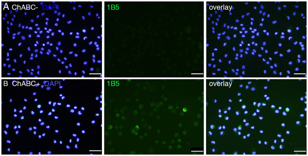
Fig 2. Immunocytochemical staining of CS stub in expanded SCE-UCBCs.
Representative photomicrographs of the staining of 1B5 (unsulfated CS stub) monoclonal antibody in expanded SCE-UCBCs. Immunoreactivity was only observed in the cells pre-digested with Chase ABC (B), and not in the cells not digested with Chase ABC (A, see Methods). blue, DAPI; green, 1B5 antibody. Bar, 50 μm.

